-
Gallery of Images:

-
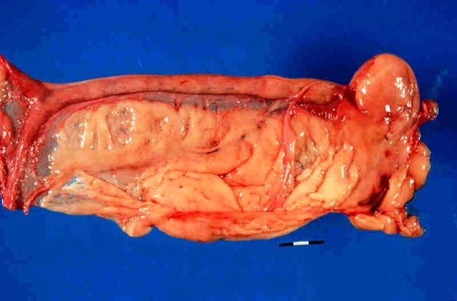
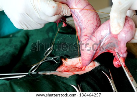
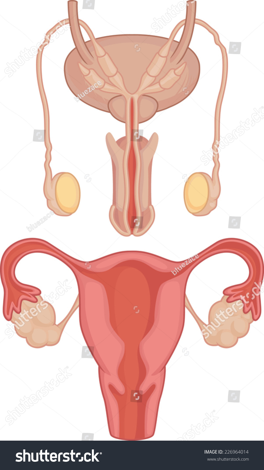
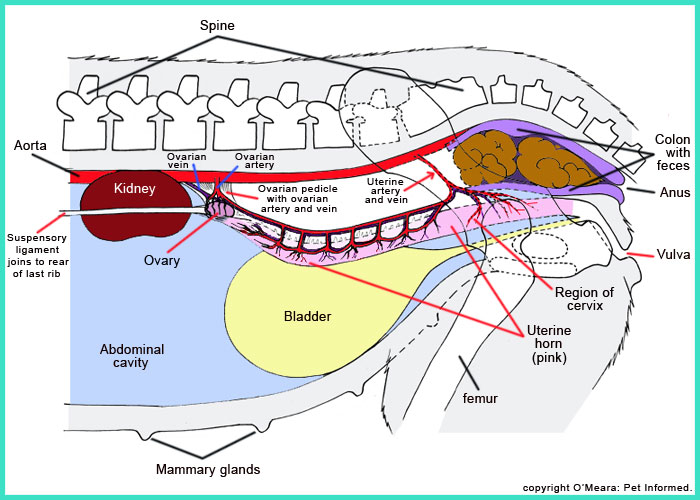
Pyometra is often the result of hormonal changes in the reproductive tract. Following oestrus (heat) in the dog, progesterone levels remain elevated for eight to ten weeks and thicken the lining of the uterus in preparation for pregnancy. Dunn and Foster (1977) reports on a dog with peritonitis secondary to perforation of the uterus in a dog with pyometra. E coli was cultured from the uterus. Kitzman (1978) reported a rupture of the uterus and a dead puppy was found in the peritoneal cavity. Some canine anatomical names may be familiar to you dogs have elbows and ears and eyes but other names may be downright foreign. Many anatomical terms used to describe parts of a dog are similar to the ones used for horses. Start studying Dog Anatomy Genital Organ Functions of the Male Dog. Learn vocabulary, terms, and more with flashcards, games, and other study tools. In this image, you will find Fimbriae, Ovary, Cervix, Vagina, Perimetrium, Myometrium, Endometrium in it. We are pleased to provide you with the picture named Uterus and uterine tubes. We hope this picture Uterus and uterine tubes can help you and satisfies your requirements. com found Uterus and uterine tubes from plenty anatomical pictures on. A dog's uterus is a bit different than a human's. The uterus is Yshaped, with the body being the main part of the uterus and each arm of the Y called horns. The ovaries are at the end of each horn, and puppies develop in the horns. Urogenital System of the Dog The pictures in this section are reprinted with permission by the copyright owner, Hill's Pet Nutrition, from the Atlas of Veterinary Clinical Anatomy. These illustrations should not be downloaded, printed or copied except for personal, noncommercial use. A woman can have two each of the uterus, cervix, and vagina. Another case is when a woman has uterus duplex bicollis two each of the uterus and cervix, but only one vagina. Ultrasound, vaginoscopy (insertion of a scope into the vagina to view the internal anatomy), and hysterosalpingogram (HSG, an imaging procedure that. The ligaments of the female reproductive tract can be divided into three categories: Broad ligament a sheet of peritoneum, associated with both the uterus and ovaries. Uterine ligaments ligaments primarily associated with the uterus. The uterus or womb is part of the reproductive system of the female body. The uterus is the place a baby grows for nine months during pregnancy. It is a pearshaped organ inside a woman. The dogs uterus plays very important roles in the intact female dogs body. This reproductive organ is similar in many ways to the uterus in women, but its also different in many other ways. The reproductive organs of a male dog File: Anatomy and physiology of animals Diagram summarizing the functions of the male reproductive organs. 3 Diagram summarizing the functions of the male reproductive organs An introduction to the anatomy of the uterine cervix cervix is the lower fibromuscular portion of the uterus. It is cylindrical or conical in shape, and measures 3 to 4 cm in length, and 2. It is supported by An introduction to the anatomy of the uterine cervix. Let's begin by going over the reproductive anatomy of dogs and pointing out some unique features. One unique feature of male dogs as compared to many other mammals is the presence of only one accessory sex gland. The female genital tract includes the vulva, vagina, cervix, uterus, oviducts, and ovaries. The mammary glands, found on the chest and abdomen, are also part of the reproductive system. The abdominal aorta gives rise to the external iliac a. gives rise to the pudendoepigastric trunk, which diverts into external pudendal a. The anatomy of the uterus consists of the following 3 tissue layers (see the following image): The inner layer, called the endometrium, is the most active layer and responds to cyclic ovarian hormone changes; the endometrium is highly specialized. The female reproductive system is designed to carry out several functions. It produces the female egg cells necessary for reproduction, called the ova or oocytes. The uterus is an organ of the female reproductive system. Its shaped like an upsidedown pear and has thick walls. The uteruss main function is to house and nourish a fetus until its. Anatomy is a branch of biology and medicine that studies the morphology and structure of living organisms. And female dog anatomy aims at making a study of all parts of the female dogs body. The detailed structure depends on a lot of factors such as the dog breed, age, and weight. The uterus and ovaries are the most vital organs of the female reproductive system. These organs work together to produce female sex hormones, produce and develop ova (egg cells), and support the development of a fetus during pregnancy. The female dogs reproductive tract consists of the female genital organs including the ovaries, uterus, vagina, vulva, and mammary glands. Where Is the Female Canine Reproductive Tract Located? The reproductive organs are located in the abdomen, and the mammary glands are located in two rows along the outside of the abdomen, running from the. The broad ligament of the uterus is the wide fold of peritoneum that connects the sides of the uterus to the walls and floor of the pelvis LAB 17 Introduction. Abdominal Viscera Nerves Peritoneal Structures (Guide to the Dissection of the Dog, 8th ed. ) broad ligament of the uterus (connecting peritoneum): The minor duodenal papilla, which receives the accessory pancreatic duct, is generally present in the dog but present in only a minority (20) of cats. The uterus is a secondary sex organ. Secondary sex organs are components of the reproductive tract that mature during puberty under the influence of sex hormones produced from primary sex organs (the ovaries in females and the testes in males). The female genital tract includes the vulva, vagina, cervix, uterus, oviducts, and ovaries. The mammary glands, found on the chest and abdomen, are also part of the reproductive system. Your dog's reproductive system consists of a vagina, cervix, uterus, oviducts and ovaries. Her ovaries produce unfertilized eggs and the hormones associated with oestrus and pregnancy. The eggs travel from the ovaries to her oviducts, where the eggs are fertilized by sperm. The uterus (from Latin uterus, plural uteri) or womb is a major female hormoneresponsive secondary sex organ of the reproductive system in humans and most other mammals. In the human, the lower end of the uterus, the cervix, opens into the vagina, while the upper end, the fundus, is. A female dog's reproductive system involves the uterus, the cervix, the oviducts, the ovaries, and the vagina. The ovaries are the organs that are responsible for. Dr Greg shows what is involved in a pyometra diagnosis and surgery in a nonspayed female dog. The gross anatomy of a dog and a human consists of a set of biological systemsthe skeletal and integumentary, digestive, muscular, lymphatic and endocrine, cardiovascular, respiratory, nervous, urinary, male and female reproductive and urinary systems. The ovaries are the female pelvic reproductive organs that house the ova and are also responsible for the production of sex hormones. They are paired organs located on either side of the uterus within the broad ligament below the uterine (fallopian) tubes. The ovary is within the ovarian fossa, a space that is bound by the external iliac vessels, obliterated umbilical artery, and the ureter. Vagina and Vestibule Anatomy Physiology. Extends from the external ostium of the uterus to the entrance of the urethra. During the dog's 'tie the bulbs are erect and press against the penis, caudal to the enlarged bulb of the glans. The cervix of the uterus is the tapered inferior region of the uterus. Its name, cervix, comes from the Latin word meaning neck due to its role as the narrow connection between the larger body of the uterus above the vagina below. The cervix plays vital roles in the control of movement into. uterus ultrasound education showing how to, scanning protocol, normal anatomy, anatomic variants, myometrium, endometrium, bicornuate, cervix, retroverted Please add to GA account UA with Manage Users and Edit permissions date Aug 10, 2017. Anatomy Of Human Organs Annahamilton. Anatomy Of Human Organs Annahamilton. me Anatomy Of Dog Canine Tooth. Anatomy Of The Brain Emedicine. Uterus Anatomy The female dog's uterus is not the same as a human uterus, but is shaped as a Y rather than a pear. The base of the uterus is the cervix, which remains closed as much as possible to prevent foreign matter or infection from entering the uterus. The uterus varies considerably in size, shape and weight depending on the status of parturition and estrogenic stimulation. The uterus is a fibromuscular organ that can be divided into the upper muscular uterine corpus and the lower fibrous cervix, which extends into the vagina. Reproductive Tract Anatomy of the Bitch Uterus During Estrus. Documents Similar To Dog Reproductive Anatomy and Physiology. New York Special contents; Categories list; Topics list; Sitemap 3D Canine Anatomy software. Biospheras innovative 3D software demonstrates the various internal systems of a virtual German Shepherd dog. The software is designed with students, teachers, veterinary clinics and pet shops in mind and allows the dogs internal systems to be viewed layer by layer. The Uterus is the organ of pregnancy as this is where implantation and development of the feotus occurs. The Uterus is the reproductive organ with the most species variations. These variations occur in both the anatomical types of uterus as well as the uterine horn appearance and endometrial linings. In this lecture we will discuss basic reproduction of the dog. Let's begin by going over the reproductive anatomy of dogs and pointing out some unique features. This is the result of the loss of red blood cells by leaking of the blood vessels in the uterus as it prepares for mating. Anatomy Of Dog Uterus In this website we provide best clipart about Anatomy Of Dog Uterus that we have collected from any source about clipart. Find Your clipart here for your refrence, and of course what we provide is the most up to date of clipart for Anatomy Of Dog Uterus. Anatomy Of Dog Uterus In this website we provide best clipart about Anatomy Of Dog Uterus that we have collected from any source about clipart. Find Your clipart here for your refrence, and of course what we provide is the most up to date of clipart for Anatomy Of Dog Uterus. Prolapsed uterus is a condition that generally occurs in female cats. However, this disease is seen in a few female dogs and is known as a female reproductive disorder. Uterine prolapse occurs when the uterus protrudes through the dogs vagina, particularly after whelping..
-
Related Images: